 Best pictures from the world of medicine
[ 2009-10-21 14:04 ]
11 老鼠肝臟細胞
這張顯微圖片顯示的是老鼠肝臟的內部結構,有助于理解該復雜器官。呈正弦曲線的血管是圖中遍及肝臟內部的粉紅色結構,血管中包含著血紅細胞和庫普弗細胞,它們是肝臟內部的巨噬細胞。肝細胞是圖中褐色部分,圍繞著正弦曲線血管排列著。
膽汁被分泌進小管之中,圖中以綠色管道顯示,它們是肝細胞之間擴大的細胞間隙,膽汁在其中流向小腸。
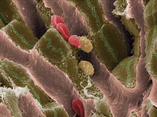
This image shows the internal structure of a mouse liver, helping to understand this complex organ. Sinusoids – blood vessels – can be seen as pink structures running through the tissue. These contain red blood cells and Kupffer cells, which are specialist macrophages of the liver. Liver cells, or hepatocytes, shown in brown, are arranged in plates surrounding the sinusoids. Bile is secreted into the passages known as canaliculi, shown as green channels. They are dilated intercellular spaces between hepatocytes, and bile flows through them en route to the small intestine. (Image: Jackie Lewin) [NewScientist]
|