 Best pictures from the world of medicine
[ 2009-10-21 14:04 ]
13 老鼠小腸內壁3D結構
如圖所示,這是使用多光子熒光方法呈現的老鼠小腸的內壁3D結構,在小腸內壁的指狀長茸毛可增大小腸內壁表面積,因而有助于消化。通過結合多張圖片的觀測,保羅-阿普爾頓(Paul Appleton)和他的同事們得以調查結腸癌導致的小腸內病變。照片是由保羅-阿普爾頓(Paul Appleton)提供。
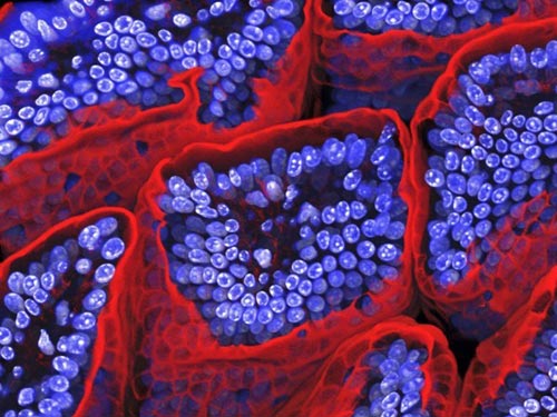
This 3D reconstruction of the lining of a mouse's small intestine was made using multiphoton fluorescent methods. Villi are small finger-like projections in the lining that increase surface area and so assist digestion. By combining stacked images, Paul Appleton and his colleagues were able to investigate the changes that colon cancer causes in the intestines. (Image: Paul Appleton) [NewScientist]
|